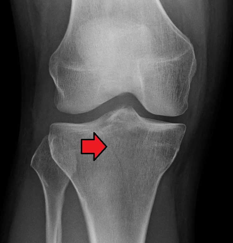
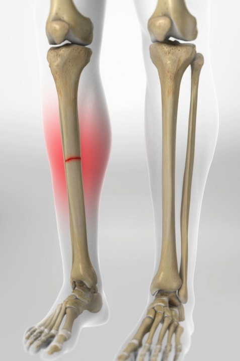
The tibia or shin is an important bone in the leg that connects the knee to the ankle.. A tibia fracture is a break in the continuity of the shin (tibia).
Proximal tibia fractures: a Tibial fracture proximal is a tear in the upper part of the shin or tibia. Proximal tibia fractures may or may not affect the knee joint. Fractures entering the knee joint can cause joint imperfections, uneven joint surfaces and improper leg alignment. This can lead to joint instability., arthritis and loss of movement. These fractures are caused by stress or trauma or in a bone already compromised by disease, like cancer or infection. Proximal tibial fractures can lead to injury to surrounding soft tissues, including skin, the muscles, the nerves, blood vessels and ligaments.
Symptoms of tibia fracture include painful weight bearing movements, tension around the knee, limitation of movement and deformity around the knee. In some people, impaired blood supply as a result of the fracture can lead to a pale or cold foot. Patients may also experience numbness or a feeling of “tingle” in the foot as a result of an associated nerve injury.
Fracturas de la tibia proximal (espinilla)
Una fractura o rotura en la tibia justo debajo de la rodilla se llama fractura de tibia proximal. La tibia proximal es la parte superior del hueso donde se ensancha para ayudar a formar la articulación de la rodilla.
Además del hueso roto, los tejidos blandos (piel, músculos, nervios, vasos sanguíneos y ligamentos) pueden lesionarse en el momento de la fractura. Tanto el hueso roto como las lesiones de tejidos blandos deben tratarse juntos. En muchos casos, se requiere cirugía para restaurar la fuerza, el movimiento y la estabilidad de la pierna y reducir el riesgo de artritis.
La rodilla es la articulación que soporta peso más grande del cuerpo. Tres huesos se unen para formar la articulación de la rodilla: el fémur (hueso del muslo), la tibia (espinilla) y la rótula (rótula). Los ligamentos y los tendones actúan como cuerdas fuertes para mantener unidos los huesos. También funcionan como restricciones, lo que permite algunos tipos de movimientos de rodilla y otros no. Además, la forma en que se forman los extremos de los huesos ayuda a mantener la rodilla correctamente alineada.
Hay varios tipos de fracturas de tibia proximal. El hueso puede romperse en línea recta (fractura transversal) o en muchos pedazos (fractura conminuta).
A veces, estas fracturas se extienden hasta la articulación de la rodilla y separan la superficie del hueso en unas pocas (o muchas) partes. Estos tipos de fracturas se denominan fracturas intraarticulares o de meseta tibial.
La superficie superior de la tibia (la meseta tibial) está hecha de hueso esponjoso, que tiene una apariencia de “panal” y es más suave que el hueso más grueso que se encuentra debajo de la tibia. Las fracturas que involucran la meseta tibial ocurren cuando una fuerza empuja el extremo inferior del hueso del muslo (fémur) hacia el hueso blando de la meseta tibial, similar a un punzón. El impacto a menudo hace que el hueso esponjoso se comprima y quede hundido, como si fuera un trozo de espuma de poliestireno sobre el que se haya pisado.
Este daño a la superficie del hueso puede resultar en una alineación incorrecta de las extremidades y, con el tiempo, puede contribuir a la artritis, inestabilidad y pérdida de movimiento.
Las fracturas de tibia proximal pueden cerrarse, lo que significa que la piel está intacta, o abrirse. Una fractura abierta es cuando un hueso se rompe de tal manera que los fragmentos de hueso sobresalen a través de la piel o una herida penetra hasta el hueso roto. Las fracturas abiertas a menudo implican mucho más daño a los músculos, tendones y ligamentos circundantes. Tienen un mayor riesgo de problemas como infecciones y tardan más en sanar.


The proximal tibia is the upper part of the tibia that connects to the knee joint. Proximal tibia fractures are fairly common injuries to the lower leg.. They can be the result of low-energy injuries or a high-energy injury, ranging from slip and falls to major car accidents.
Because the blood vessels, ligaments, muscles, nerves and skin can be injured simultaneously during this type of fracture, it is crucial that an orthopedic specialist evaluate any soft tissue damage to properly treat the fracture. Proximal tibial fractures must be properly identified and diagnosed to manage the injury and restore normal range of motion., stability and strength of the limbs. Proper and simultaneous management of such fractures and any accompanying soft tissue injuries can also reduce the likelihood of arthritis later in life..
Since the knee supports more weight than any other joint and is responsible for a wide range of movements, injuries to the bones and ligaments that connect them can be quite serious. The muscles, Ligaments and other soft tissues play an important role in the stability of the knee joint, but not as much as the bones of the lower limbs, in particular the bone that forms the tibial plateau. The proximal tibia is not as thick as the tibial shaft, so it is more easily injured.
There are two distinct internal areas of the proximal tibia: the medial condyle and the lateral condyle. There are also two smooth articular facets on the upper surface of the proximal tibia, that articulate with the condyles of the femur in the central part and support the meniscus of the knee joint in the peripheral part.
The spine of the tibia, also known as intercondylar eminence, lies between the articular facets. This is protected by a tubercle on either side and marked by depressions where the menisci and posterior cruciate ligaments connect in the front and rear.. There are several surfaces of the proximal tibia:
Proximal tibia fractures can be more or less severe depending on whether the bone is partially or totally broken and whether the fracture is open or closed.:
A proximal tibia fracture, or any fracture for that matter, can be classified into the following categories based on various other factors:
Tibia fractures are common injuries. The subcutaneous nature of the tibia makes it more prone to open injuries. The musculature of the lower leg is divided into four compartments separated by fascial tissue. Radiographs are essential in the initial evaluation of fractures. In the case of injury or fracture of the lower limb, fascial tissue may need to be released through fasciotomies to avoid sequelae of compartment syndrome. Treatment methods may be nonsurgical for minimally displaced fractures., although surgical fixation is preferred for open and displaced fractures.
The tibia is the main bone of the lower leg and forms what is more commonly known as the shin.
Expands at its proximal and distal ends; articulating at the knee and ankle joints respectively. The tibia is the second largest bone in the body and is a key structure to bear weight .
The proximal tibia widens with the medial and lateral condyles, that help to bear weight. The condyles form a flat surface, known as tibial plateau. This structure articulates with the femoral condyles to form the key joint of the knee joint..
Between the condyles is a region called intercondylar eminence , projecting upward on both sides as the medial and lateral intercondylar tubercles. This area is the main site of attachment of the ligaments and menisci of the knee joint.. The intercondylar tubercles of the tibia articulate with the intercondylar fossa of the femur.
The body of the tibia is shaped like prism , with three edges and three surfaces; anterior, back and side. For the sake of brevity, here only the edges are mentioned / anatomically and clinically important surfaces.
The distal end of the tibia is widens to help bear weight.
The maléolo medial is a bony projection that continues down the medial aspect of the tibia. Articulates with the tarsal bones to form part of the ankle joint. On the posterior surface of the tibia, there is a groove through which the tibialis posterior tendon passes.
Sideways is the notch of the fibula, where the fibula joins the tibia, forming the distal tibiofibular joint.
A fracture or break in the tibia just below the knee is called a proximal tibial fracture.. The proximal tibia is the top of the bone where it widens to help form the knee joint..
Besides the broken bone, soft tissues (skin, muscles, nerves, blood vessels and ligaments) may be injured at the time of the fracture. Both the broken bone and any soft tissue injury should be treated together. In many cases, surgery is required to restore strength, movement and stability of the leg and reduce the risk of arthritis.
The knee is the largest weight-bearing joint in the body. Three bones join together to form the knee joint: The femur (thigh bone), the warm (shin) and the kneecap (ball joint). Ligaments and tendons act as strong cords to hold bones together. They also work as restrictions, allowing some types of knee movements and others not. Besides, the way the ends of the bones are formed helps keep the knee in proper alignment.

There are several types of proximal tibia fractures. Also called tibial plateau fractures. The bone can break in a straight line (transverse fracture) or in many pieces (comminuted fracture).
Sometimes, These fractures extend to the knee joint and separate the surface of the bone in a few (or many) parts. These types of fractures are called intra-articular fractures..
The upper surface of the tibia (the tibial plateau) is made of cancellous bone, which has an appearance of “diaper” and is softer than the thickest bone under the tibia. Fractures involving the tibial plateau occur when a force pushes the lower end of the thigh bone (femur) towards the soft bone of the tibial plateau, similar to a punch. Impact often causes cancellous bone to compress and collapse, as if it were a piece of styrofoam that has been stepped on.
This damage to the bone surface can result in incorrect alignment of the limbs and, over time, can contribute to arthritis, instability and loss of movement.
Proximal tibial fractures can be closed, which means the skin is intact, or open up. An open fracture is when a bone is broken in such a way that the bone fragments protrude through the skin or a wound penetrates to the broken bone. Open fractures often involve much more damage to the muscles, surrounding tendons and ligaments. They are at higher risk for problems such as infections and take longer to heal.
There are several types of fractures of the upper tibia. The bone can break in a straight line (transverse fracture) or in many pieces (comminuted fracture)
Sometimes, These fractures extend to the knee joint and separate the surface of the bone in a few (or many) parts. These types of fractures are called intra-articular or tibial plateau fractures..
The upper surface of the tibia (the tibial plateau) is made of cancellous bone, which has an appearance of “diaper” and is softer than the thickest bone under the tibia.
Fractures involving the tibial plateau occur when a force pushes the lower end of the thigh bone (femur) towards the soft bone of the tibial plateau, similar to a punch. Impact often causes cancellous bone to compress and collapse, like it's a styrofoam piece that has been stepped on.
This damage to the bone surface can result in incorrect alignment of the limbs and, over time, can contribute to arthritis, instability and loss of movement.
Proximal tibial fractures can be closed, which means the skin is intact, or open up. An open fracture is when a bone is broken in such a way that the bone fragments protrude through the skin or a wound penetrates to the broken bone. Open fractures often involve much more damage to the muscles, surrounding tendons and ligaments. They are at higher risk for problems such as infections and take longer to heal.
A fracture of the upper part of the tibia can occur from stress (minor breaks due to unusual excessive activity) or for a bone already compromised (as in cancer or infection). The majority, however, are the result of trauma (injury).
Young people often experience these fractures as a result of a high-energy injury., like a fall from a considerable height, sports-related injuries and car accidents.
Older people with poorer quality bones often only require a low-energy injury (fall from a standing position) to create these fractures.
Symptoms include:
If you have these symptoms after an injury, go to the nearest hospital emergency room for an evaluation.
Meet our Portfolio of Services and Specialties focused on Orthopedics and Traumatology of the Shoulder and Knee.

Orthopedist and Traumatologist
Schedule your Appointment with him Dr. Danilo Velandia to be able to provide you with information and guidance about our Procedures. All the necessary information will be explained to you from: how to prepare for surgery, what will happen to you at the Clinic or Hospital, how you should take care of yourself at the end of the Procedure, as well as the changes that must occur in your lifestyle.
I broke my left rotator cuff while falling. I tried to put up with it for almost a year with medications, physical therapy and basically it didn't help . I was scheduled for surgery elsewhere. I did not like the atmosphere, so I looked for another opinion. I am happy to have done it !! all nursing staff, ambulatory surgical units, postoperative care and now the final results: I have no pain I have a full range of motion, I have strength and continue to develop it while following a very detailed therapy regimen. I can only recommend Dr.. Danilo Velandia and elite staff, professional, personal, warm and friendly.

I am so impressed and grateful for the care that Dr.. Danilo, that every time I get a chance to give a positive review, I can not resist! The level of care he provides far exceeds that of any other doctor I have ever seen. I am so impressed. He patiently explained every detail of the treatment plan for my broken Knee to me and really took the time to listen and answer all the questions I had.. If I ever have to refer someone I know to an orthopedic surgeon, I will insist that they see Dr.. Danilo Velandia. There is no one else I would turn to if I needed one again. Thank you Dr. Danilo for the great care you have given me!

I have seen Dr.. Danilo Velandia a couple of times in Bogotá and I'll be back, it's been great so far. No pressure to rush to surgery, great advantage. Gives good information and options from the first day to the surgery process. All staff are friendly and helpful highly qualified, He has been the most proactive and helpful medical person I have dealt with and I am really very happy. Your diligence has made sure you get the help you need, and it even helped me avoid forgetting things. Thank you very much indeed! I am very happy with my visits with him. Highly recommended Dr. Danilo Velandia and his team of professionals!

My experience as a patient of Dr.. Daniel has been extremely positive. Performed surgery on my right shoulder, completely relieving my symptoms. He patiently listened to my concerns, answered my many questions and provided insightful information, but not too technical. The doctor. Danilo made himself available both before and after my surgery to address my pre-surgery nerves and my post-surgery expectations.. He was always professional, patient and attentive. I feel very lucky that when I could no longer suffer with the symptoms, my husband, who recommended me to Dr.. Danilo Velandia.

After playing competitive soccer for several years, I broke my anterior cruciate ligament and meniscus. I was very nervous about the long process of surgery and recovery really is a life changing process, I thought I would lose everything since sport is my life and my passion, but Dr.. Danilo Velandia was very helpful, informative and reassuring, he really knew how to handle the situation. My surgery went smoothly and my recovery went according to plan. The doctor. Danilo was present every step of the way and showed his medical competence, as well as his natural compassion, Thank you very much and very grateful, I have been able to continue with my day to day.

***We Promise, no spam!
Doctor Danilo Velandia León Specialist with experience in Orthopedics and Traumatology, Knee Surgery and Shoulder Surgery, which includes prevention, diagnosis, surgical and non-surgical treatment and monitoring of all conditions of the musculoskeletal system and its associated structures (bones, joints, ligaments, tendons, muscles and nerves)
Our Services are designed and developed to treat congenital pathologies, deformities, sports injuries, degenerative lesions and osteoarthritis. Advanced surgical and non-surgical techniques. Arthroscopy, osteotomies and joint replacements. Successful Recovery and Rehabilitation to support the successful recovery and rehabilitation of our Patients.
Orthopedic surgeon in Bogotá © 2021, Doctor Danilo Velandia León – Orthopedist and Traumatologist. Web jobs All Rights Reserved.